Centro Dental Las Americas provides digital X-rays in Hyattsville, MD to help diagnose cavities, gum disease, infections, and other oral health concerns with speed and accuracy. This page explains how digital dental radiography works, what images may be taken, safety considerations, and what you can expect during your visit.
Digital X-Rays Explained
Digital X-rays use electronic sensors instead of traditional film to capture detailed images of your teeth, roots, and jaw. The images appear on a screen within seconds, allowing the dental team to enhance contrast, zoom in, and compare past and present views. This clarity supports precise diagnosis and treatment planning.
These images help identify issues that are difficult or impossible to see during a visual exam, such as cavities between teeth, bone loss around roots, impacted teeth, infections, and changes beneath restorations. Because the data is digital, images can be securely stored, shared with specialists when needed, and tracked over time to monitor your oral health.
Common Types of Images
- Bitewing images show the upper and lower back teeth and are useful for finding cavities between teeth and checking bone levels.
- Periapical images focus on one or two teeth from crown to root tip to evaluate infections, fractures, and root concerns.
- Panoramic images capture the entire mouth and jaw in one view to assess wisdom teeth, jaw joints, cysts, or impacted teeth.
- Occlusal images show a broad area of the mouth and are sometimes used to evaluate developing teeth in children.
- In select cases, a 3D cone beam scan (CBCT) may be recommended for complex planning, such as implant evaluation.
Benefits of Digital X-Rays
- Reduced radiation compared with traditional film, following the ALARA principle (As Low As Reasonably Achievable).
- Immediate images that speed up diagnosis and reduce time in the chair.
- High-resolution detail that helps reveal small cavities and early bone changes.
- Easy image sharing for second opinions or referrals when necessary.
- Eco-friendly workflow that avoids chemical film processing.
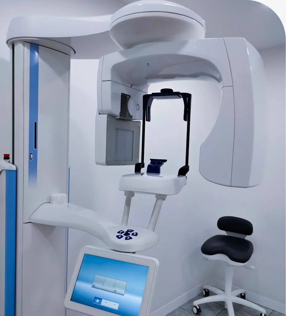
How Digital X-Rays Work
- Preparation: You will wear a protective lead apron, and often a thyroid collar, for added shielding.
- Sensor placement: A small digital sensor or plate is positioned in your mouth. A bite tab helps hold it steady.
- Image capture: The X-ray unit briefly activates while you remain still. The exposure lasts a fraction of a second.
- On-screen review: The image appears on a monitor for immediate review, where brightness and contrast can be adjusted.
- Discussion: The dentist reviews findings with you and explains any recommended next steps.
What to Expect
Most digital X-rays are quick and comfortable. You may feel slight pressure from the sensor against your cheeks or tongue, which eases as you adjust. If you have a strong gag reflex, breathing through your nose and focusing on a fixed point can help. Children and adults with smaller mouths may benefit from child-sized sensors.
Frequency depends on your age, oral health, cavity risk, and symptoms. Many healthy adults receive bitewings every one to two years, while higher-risk patients may need them more often. Panoramic images are taken when broader evaluation is needed, such as during orthodontic planning or wisdom tooth assessment. Your dentist tailors the schedule to your needs.
Safety is a top priority. Digital sensors require far less radiation than film, and shielding further reduces exposure. Always inform the team if you are pregnant or think you could be. Necessary dental X-rays are considered safe during pregnancy with appropriate protection, but non-urgent images may be postponed until after delivery.
Related Services
Looking for related care at our Hyattsville, MD dental office? Explore these treatments:
Frequently Asked Questions About Digital X-Rays
Yes. Digital imaging uses low radiation doses, combined with protective aprons and careful technique. The benefit of early detection outweighs the small exposure for most patients.
It varies. Your dentist considers your cavity risk, history of dental work, gum health, and symptoms. Many adults need bitewings every one to two years, while others may need them more or less often.
They can show decay between teeth, infections at the root, bone loss from gum disease, impacted teeth, and problems under existing fillings or crowns. They also help plan treatments like implants or root canals.
Tell the dental team as early as possible. Urgent X-Rays can be taken with shielding, but routine images are often delayed until after pregnancy when appropriate.
Often, yes. Children benefit from X-rays to monitor tooth development and detect decay between teeth. Child-sized sensors and careful techniques keep exposure low.
Digital X-rays offer lower radiation, faster results, and clearer images that can be enhanced on screen. They also eliminate chemical processing and streamline record sharing.