Panoramic X-rays provide a single, comprehensive image of your teeth, jaws, and surrounding structures. Panoramic X-rays in Hyattsville, Maryland, help our dentist evaluate areas that standard bitewing images cannot capture, offering a wide view that supports accurate diagnosis and treatment planning with minimal discomfort.
Panoramic X-Rays Explained
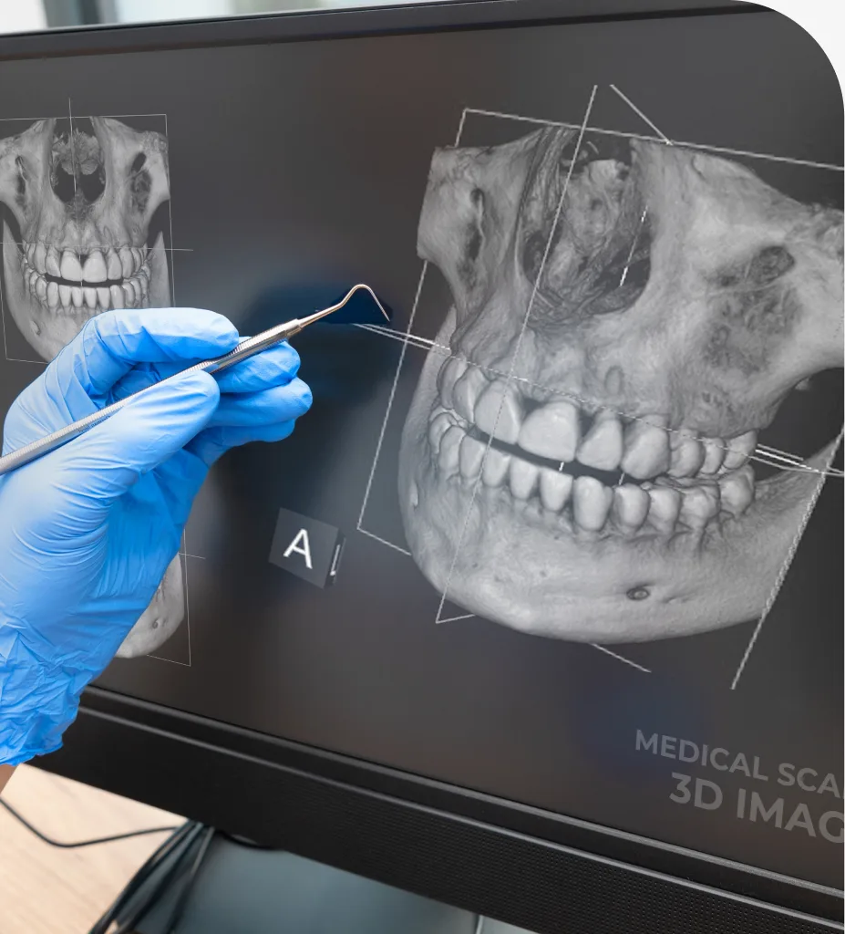
A panoramic dental X-ray is a two-dimensional image that shows the entire mouth in one scan. Unlike small, close-up X-rays placed inside the mouth, this extraoral image is taken by a machine that rotates around your head. The result is a broad snapshot of the upper and lower jaws, all teeth, temporomandibular joints (TMJ), sinuses, and parts of the facial bones.
This imaging is useful at many stages of care. Common uses of panoramic X-rays in dentistry include evaluating wisdom teeth position, assessing growth and development, screening for cysts or tumors, checking jaw joint health, planning dental implants, and reviewing bone levels. While it is not the best tool for small cavity detection, it pairs well with other images to build a complete picture of oral health.
Why Consider Panoramic Dental Imaging?
Patients often ask what the benefits of panoramic dental imaging are and how it compares to other options. Key advantages include:
- Broad coverage for a full-mouth overview, including teeth, jaws, and sinuses.
- Convenient experience with no sensors inside the mouth, helpful for a strong gag reflex.
- Efficient imaging that is typically completed in seconds and reviewed the same day.
- Valuable planning for implants, extractions, orthodontics, and other treatments.
- Low radiation dose that is comparable to a small amount of natural background exposure.
- Versatility to help identify issues that may not appear on bitewing or periapical images.
If you are comparing a panoramic X-ray vs. bitewing images, remember each serves a different purpose. Bitewings are best for detecting small cavities between teeth, while a panoramic radiograph offers the “big picture” view of the jaws and structures beyond the teeth.
The Panoramic X-Ray Process
The process is straightforward and designed for comfort. Here is how a typical visit goes:
- Preparation: You will remove glasses, earrings, or metal objects and wear a protective apron.
- Positioning: You will stand or sit with your chin on a rest and gently bite on a small guide to steady your jaw.
- Imaging: The machine’s arm rotates around your head for about 10 to 20 seconds as it captures the image.
- Review: The digital image appears on a screen, and your dentist reviews it with you to discuss findings and next steps.
What To Expect Before And After
Most patients find panoramic X-rays quick and easy. You can breathe normally, and no sensors go inside your mouth. The radiation exposure is low and carefully controlled, and modern equipment uses settings tailored to your size and needs. In many cases, the dose is similar to the amount of background radiation you receive over a few days.
Results are available right away. If the panoramic image raises questions about a specific tooth or area, your dentist may supplement it with a small, close-up X-ray or 3D scan for fine detail. This layered approach helps answer “what is a panoramic dental X-ray used for” and “how does a panoramic X-ray work” in real-world care: it is the broad view that guides targeted, precise imaging when needed.
Patients can expect a calm visit with clear explanations. If you are pregnant or think you might be, alert the dental team in advance so appropriate precautions and timing can be discussed.
Related Services
Looking for related care at our Hyattsville, MD dental office? Explore these treatments:
Frequently Asked Questions About Panoramic X-Rays
Panoramic images can show impacted teeth, jaw fractures, cysts or tumors, sinus issues, TMJ changes, bone loss patterns, and tooth development. They are also useful for planning implants and extractions.
Yes. Dental X-ray safety standards are strict, and equipment uses the lowest radiation dose needed for a clear image. Protective aprons and careful positioning further reduce exposure.
A panoramic X-ray captures the entire mouth at once and is ideal for an overall view of the jaws and structures. Bitewings focus on the crowns of the back teeth and are better for spotting small cavities between teeth.
The scan itself usually takes less than 20 seconds and is painless. You will be asked to hold still and bite gently on a guide while the machine rotates around your head.
Non-urgent imaging is often postponed during pregnancy. If a panoramic X-ray is necessary, shielding and strict safety protocols are used. Always inform your dentist if you are or may be pregnant.
Frequency depends on your dental needs, age, and medical history. Your dentist will recommend timing based on screening goals, treatment planning, or monitoring specific conditions.